Facial Nerve Palsy
Red flags
Facial nerve palsy associated with severe otalgia may be a result of mastoiditis or necrotising otitis externa.
Facial palsy with parotid swelling in an elderly patient may represent parotid malignancy.
Facial palsy associated with trauma may require urgent decompression.
Other associated neurological symptoms may suggest a central cause such as stroke.
Why is this important?
Facial palsy is a common presentation to ENT, and is commonly referred to as “Bell’s Palsy”. This term is often used inaccurately, as it refers to idiopathic facial palsy only.
It is essential to rule out other conditions (eg stroke), and treat appropriately. Make sure that the patient truly has an isolated lower-motor-neurone VII palsy (ie the forehead is paralysed). If the forehead is spared, or there are other associated neurological symptoms, a central cause will be responsible.
The integrity of the facial nerve can be interrupted at any point in its course from the brainstem, to the inner ear, middle ear, parotid gland, and finally the facial musculature.
When to involve the ENT registrar
All cases with an otological cause (mastoiditis, necrotising otitis externa) should be discussed urgently, as the patient may require urgent treatment.
Traumatic facial palsy may require urgent exploration (but remember that serious intracranial injury must be excluded first).
All cases with a suspected malignant cause should be discussed with a senior during working hours.
Who to admit
Patients with possible necrotising otitis externa (often elderly diabetic patients with severe, unrelenting otalgia, associated with facial droop), must be admitted for IV antibiotics and an urgent CT of the temporal bones.
Patients with facial palsy associated with mastoiditis must be admitted for IV antibiotics and an urgent CT of the temporal bones.
Patients with a history of head injury should be admitted under the team normally dealing with head trauma, but also seen urgently by the ENT team.
Some units will admit patients with facial palsy associated with acute otitis media for IV antibiotics and urgent myringotomy and grommet insertion.
Assessment and recognition
Clinical signs
The patient may complain of facial weakness or droop, or it may be noticed by healthcare professionals or relatives. The weakness may cause asymmetry at rest, or only on movement of the face. Inability to close the eye may result in conjunctivitis and chemosis.
Because both the nerve to stapedius and the chorda tympani are branches of the facial nerve, patients may present with hyperacusis or loss of taste sensation in the anterior 2/3 of the tongue, depending on the site and type of lesion.
Careful neurological examination is essential to establish that there is isolated VIIth nerve palsy. Patients with other cranial nerve lesions may have a central cause, or potentially a skull base tumour – MRI and a neurological referral may be required.
Facial paralysis is typically graded by severity according to the House-Brackmann scale. This should be documented so that improvement or deterioration is easy to detect:
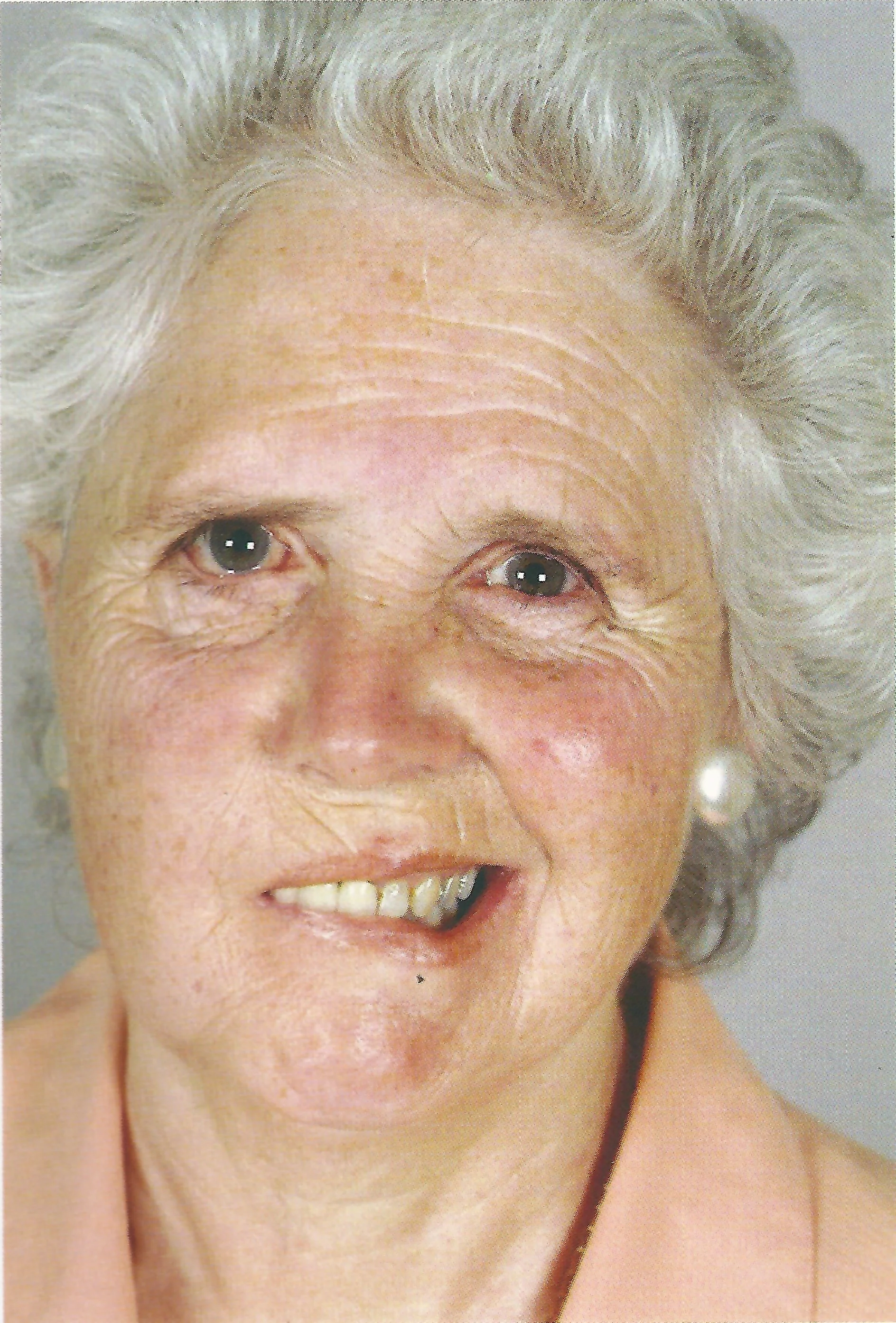
Photo - Right lower motor neurone facial palsy. Note the loss of the forehead and brow furrows as well as the nasolabial fold on the right. Similarly, the right depressor angulae oris is weak.
Grade I: Normal
Grade II: Mild asymmetry on movement
Grade III: Marked asymmetry on movement, eye closure intact
Grade IV: Marked asymmetry on movement, eye closure compromised.
Grade V: Asymmetry at rest, eye closure compromised.
Grade VI: No facial movement.
It is helpful to remember that incomplete eye closure implies Grade IV or more.
Examine the ear for otitis externa, otitis media, chronic otitis media (or cholesteatoma), or the presence of scabby vesicles, implying Ramsay-Hunt syndrome (herpes zoster oticus).
Examine the parotid for swelling (?parotid tumour). Also examine the oral cavity looking for parapharyngeal swelling.
Examine the eye, firstly to establish occlusion. If the eye does not close fully, the patient must be reviewed by an ophthalmologist and eye protection provided.
Investigations
An FBC, U+Es and CRP should be taken in all patients admitted due to an infectious cause of facial palsy.
If necrotising otitis externa or a complication of middle ear infection is suspected, or there is a history of head trauma, a CT of the temporal bones is usually necessary. Discuss this with a registrar.
An audiogram should be performed on a semi-urgent basis.
Differential diagnosis
A surgical sieve is useful for facial droop:
Idiopathic
Bell’s palsy (most common cause, a diagnosis of exclusion)
Infection
Acute otitis media +/- mastoiditis
Necrotising otitis externa
Lyme disease
Ramsay-Hunt Syndrome (Herpes zoster oticus)
Trauma
Temporal bone fracture
Surgery to middle ear or parotid
Neoplastic
Parotid carcinoma
Acoustic neuroma (patient will usually have unilateral hearing loss)
Neurological
Stroke
Guillain-Barré syndrome
Sarcoidosis
Multiple sclerosis
Immediate and overnight management
Incomplete eye closure
The eye must be protected if the patient cannot close it fully. Artificial tears (e.g. Viscotears) should be used at least QDS, and Lacrilube ointment at night. The eye should be taped closed at night or a patch worn. The patient should be reviewed by an ophthalmologist if this is the case.
Bell’s Palsy
If after careful examination no cause can be found, the diagnosis is Bell’s Palsy. This is thought to be of occult viral origin in many cases, and so most patients are prescribed high-dose oral prednisolone (up to 1 mg/kg OD for one week, then reducing), and oral aciclovir. Some pharmacists recommend gastric protection. Prednisolone slightly increases recovery rates if given within 72 hours of onset, whereas no good evidence exists for antivirals.
As for Bell’s palsy (there is still no good evidence for antivirals although they are generally prescribed). The prognosis is worse than for Bell’s palsy.
Acute otitis media or mastoiditis
Admit the patient, start IV antibiotics and steroids, keep NBM and discuss with a senior. The patient may require grommet insertion and/or cortical mastoidectomy in mastoiditis. In relatively well patients, the palsy may be due only to a dehiscent facial nerve canal as it runs through the middle ear - and may therefore not require admission. The palsy is usually incomplete or partial, with a very good prognosis once the infection has been treated.
Necrotising otitis externa
Admit the patient, perform careful microsuction and send swab cultures. Discuss with a senior and request urgent CT of the temporal bones. Start IV antibiotics and ciprofloxacin 0.3% drops, which cover Pseudomonas (the likely pathogen).
Parotid carcinoma
The patient should be referred as an urgent outpatient to head-and-neck clinic for management. Investigations include MRI and fine-needle aspiration cytology.
Further management
Bell's Palsy
Signs of recovery within four weeks are a good prognostic indicator. Typically, without treatment about 75% of people will recovery fully. With prompt steroid treatment, this increases to about 85%. Many patients with Bell’s Palsy recover in two to three months, although it may take up to nine months.
In persistent facial palsy, the eye can be protected by implanted gold/platinum weights in the eyelid or tarsorraphy. Facial reanimation by static techniques, or dynamic free muscle transfer and cross-over nerve grafting, can also be performed.
Patients should be followed up routinely in clinic to monitor progress.
References
See the Cochrane Library for many reviews on steroids and antivirals for Bell's Palsy and Ramsay-Hunt Syndrome.
Page last reviewed: 15 December 2019