
Deep Neck Space Infections
Red Flags
- The airway disaster triad (see below) alerts you to either epiglottitis or deep neck space infections (DNSI)
- Symptoms of neck stiffness and fever may be due to meningitis, and therefore this must be included in your differential diagnosis
- DNSI may progress rapidly to stridor, indicating imminent airway obstruction
Do not be fooled by a relatively well-looking patient — rapid decompensation can occur.
ENTSHO.com Impending Airway Disaster Triad
- Rapid onset aphagia or severe dysphagia, frequently associated with a severe sore throat
- Rapid onset laryngeal voice change: hoarse, croaky, husky, or no voice
- Systemically very unwell: pyrexia, tachycardia, tachypnoea
There may be associated trismus or torticollis. Beware of any patient who develops these signs after admission — situations deteriorate rapidly. In the presence of neck trauma, any of the above should be taken very seriously. Stridor is a late sign of airway compromise.
Why Is This Important?
DNSI may need surgical drainage under general anaesthetic and admission to critical care.
If left, DNSI will cause airway obstruction. It can track down into the mediastinum, requiring cardiothoracic surgical support.
When to Involve the ENT Registrar
Immediately: DNSI is an airway emergency.
Patients with deep neck space infections can rapidly develop airway obstruction (particularly children, due to their narrow airway). Always inform your senior when you are unsure of the diagnosis, as the differential includes other emergencies like epiglottitis or meningitis.
Who to Admit
Admit all patients you suspect to have a DNSI, or who have airway disaster triad symptoms.
Assessment and Recognition
History may indicate a preceding upper respiratory tract infection, ingested foreign body, peritonsillar abscess, or dental abscess.
Cardinal symptoms include the airway disaster triad, plus:
- Severe sore throat with dys- or aphagia
- Laryngeal voice change, e.g. hoarseness or voice loss
- Systemically unwell, e.g. fever and dehydration
- Neck pain and stiffness
- In children: drooling, agitation, off feeds
The differential diagnosis includes both medical and surgical emergencies, and therefore one must have a high index of suspicion for these.
Examination findings may include:
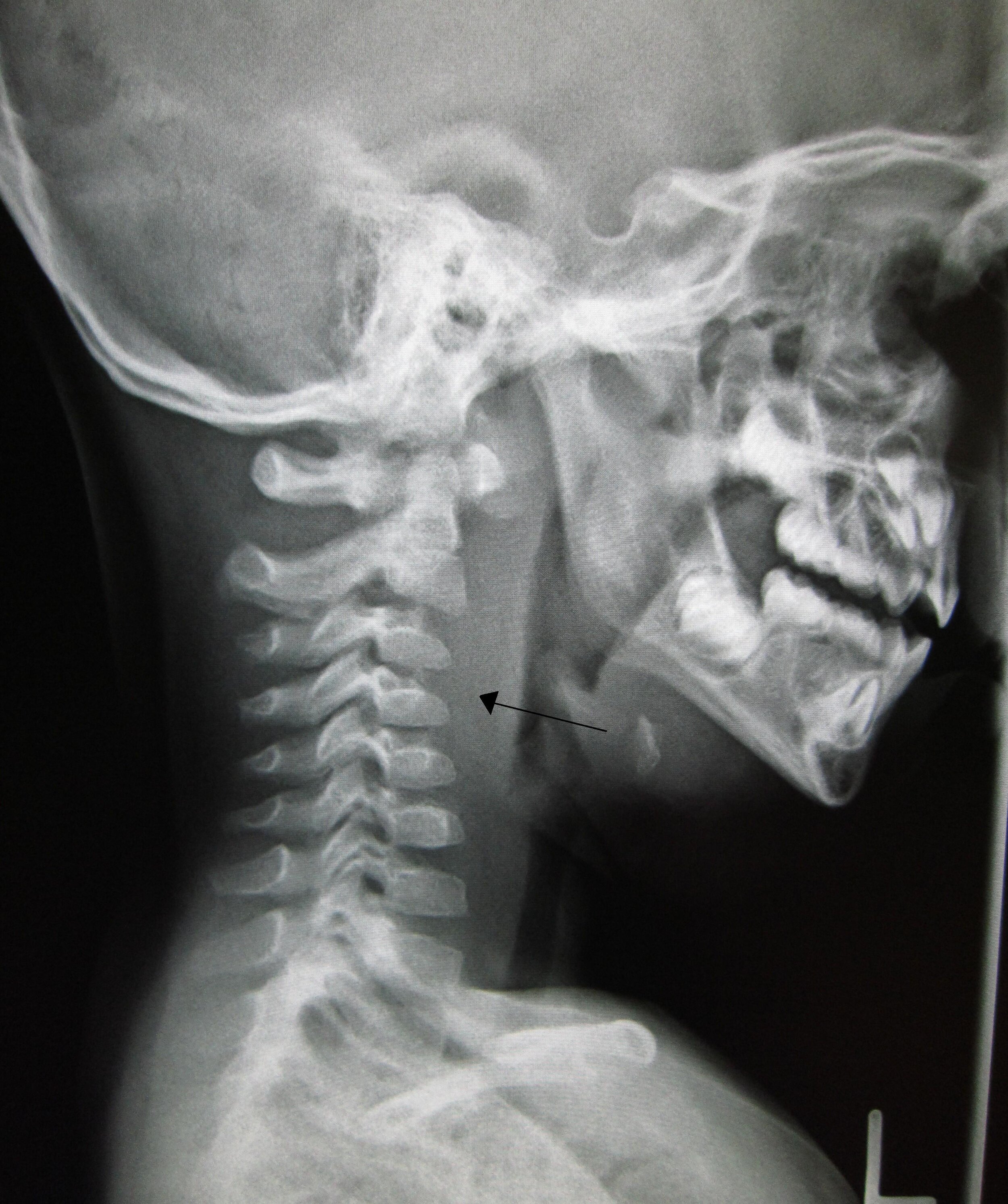
Lateral soft tissue neck X-ray showing ‘straight neck sign’ (loss of lordosis due to spasm). The arrow indicates widening of prevertebral soft tissue in a child
With thanks to James Heilman MD, Wikimedia Commons
- Stridor
- Head tilt to one side
- Trismus
- Cervical lymphadenopathy
- Unilateral smooth swelling of the posterior pharyngeal wall (the abscess is contained to one side of the midline because of the median raphe of the buccopharyngeal fascia)
- Signs of sepsis
- Drooling or agitation
- There may be concurrent tonsillitis, peritonsillitis, pharyngitis, and other upper respiratory infection, such as otitis media
Note: The general prohibition on instrumenting the mouth or performing nasendoscopy in patients with an airway problem prevents over-zealous and uncontrolled instrumentation of the base of tongue/larynx by first responders. Controlled, careful, and minimal instrumentation by trained personnel is lower risk.
Immediate and Overnight Management
Bear in mind the Sepsis Resuscitation Bundle when managing septic patients.
Follow the ABCDE algorithm. Instigate the following procedures and investigations:
- IV access + FBC (neutrophilia), CRP, U&Es, blood cultures (usually negative)
- Keep NBM until senior review and initiate IV fluids
- Initiate broad spectrum IV antibiotics and analgesia
- Humidified oxygen and back-to-back saline nebulisers
- Consider stat or PRN adrenaline nebulisers
- Flexible nasendoscopy performed by a competent endoscopist (trained SHO, ANP, or registrar) viewing the oropharynx and larynx from just beyond the soft palate; do not advance too far, as instrumenting the larynx can precipitate respiratory arrest
- Lateral soft tissue neck X-ray if safe. This may show the "straight neck sign" (Figure 1), whereby the cervical vertebral column has lost its natural lordosis. There may also be widening of the prevertebral soft tissue shadow, or a pushing forward of the airway. Very rarely, an air-fluid level may be seen in the prevertebral area.
- CT scan (ideally with contrast) will confirm the diagnosis and the extent/anatomical relations of the abscess. Make sure you have a secure airway (elective intubation or anaesthetic escort).
- Nurse upright (more than 45 degrees)
Further Management
The standard management of DNSI includes drainage, but some small abscesses will resolve with conservative treatment. Management will depend on the underlying cause, as well as the location of the abscess, for example:
- Removal of any causative foreign body that has caused a retropharyngeal abscess
- Drainage of any peritonsillar abscess, ± immediate "hot" tonsillectomy (rare), for a parapharyngeal abscess
- Drainage of dental abscess for Ludwig's angina (floor of mouth abscess)
One study found that of 162 paediatric patients with retropharyngeal abscess, 126 required surgery initially; of the 36 patients initially treated conservatively with high-dose antibiotics, 17 required surgery (1).
Drainage can be done through the mouth or through the neck (depending on the type of abscess) — consult senior colleagues.
Any sign of airway compromise may require awake intubation and/or local anaesthetic tracheostomy, which could be performed at the time of drainage.
An associated phenomenon is Lemierre's syndrome, where a DNSI is associated with an infective thrombophlebitis of the ipsilateral internal jugular vein. The causative organism is frequently Fusobacterium necrophorum; there may also be synergistic bacterial infection. It is mandatory to get a haematology opinion on anticoagulation.
Reference
- Page NC, Bauer EM, Lieu JE. Clinical features and treatment of retropharyngeal abscess in children. Otolaryngol Head Neck Surg. 2008 Mar;138(3):300-6.
Page last reviewed: 18 January 2026